Introductory
Online, live
Half day — 4 hours
Per seat, maximum 6 participants
Vea Foundations
From a DICOM series to a model you can actually explore.
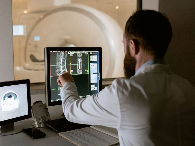
A working introduction, not a product demonstration. By the end of the session you have taken a cardiovascular dataset from import to a three-dimensional reconstruction you can navigate, annotate and show to a colleague — and you know which datasets will give you trouble before you waste an afternoon on them.
Who it is for. Clinicians, imaging specialists and researchers meeting the platform for the first time.
What we work through
- What makes a CT, MR or 3D echo series suitable for reconstruction — slice thickness, contrast timing, gating, and the compromises you can live with
- Import and reconstruction: from a DICOM series to an explorable model
- Navigating the scene — clipping planes, thresholds, isolating structures, saving and restoring views
- Keeping the reconstruction anchored to the source slices, so what you see in 3D can always be traced back to the original image
- Capturing views and recordings for a case discussion or a lecture
What is included
- Four hours live with an instructor, in a group of no more than six
- A prepared teaching dataset that stays with you
- Access to a demo environment during the session — no local installation needed
- Recording of the session
- Certificate of attendance
€690
per seat · VAT included
Dates are agreed after booking. See the booking and cancellation terms.