
Services
What we actually do
Most of it is the platform and the expertise around it. The last item on this list is the one people are usually surprised by: we build software for other organizations too.
What we offer
Patient-specific 3D visualization
Cardiovascular imaging turned into an interactive three-dimensional environment built from that patient's own data.
Immersive VR visualization
The same anatomy explored at true scale, where depth, orientation and spatial relationships are seen rather than inferred.
Cardiovascular measurement
Diameters, distances and areas in 2D and 3D, taken to a protocol that another reader can follow and reproduce.
Virtual implant simulation
Virtual positioning of a device within reconstructed anatomy, to study fit and clearance for research and teaching purposes.
Congenital heart visualization
Patient-specific review of malformed, repaired and post-surgical anatomy that conventional flat review serves poorly.
Medical imaging research
Technology and support for universities, hospitals and imaging laboratories running cardiovascular visualization studies.
Medical education
Interactive anatomy for teaching, workshops and case demonstrations, built from real datasets instead of illustrations.
PACS and imaging workflow
Connection to compatible imaging infrastructure and DICOM workflows, scoped with your IT and security teams.
Custom medical visualization software
Bespoke 3D, VR and imaging software for healthcare and research organizations, built from the same engineering base as Vea.
Custom development
Medical visualization beyond Vea
Building Vea required a specific combination of skills: medical imaging, DICOM, real-time 3D, VR, and enough clinical literacy to know which of the obvious ideas are wrong. That combination is available as a service.
Typical work is a piece of software that has to exist for a study to be possible, or a prototype that has to be convincing enough to justify the next stage of funding. We are a development partner on those projects, not a consultancy that writes a report.
Where we are useful
- Medical imaging and DICOM workflow software
- Interactive 3D and anatomical visualization
- VR and XR applications for research and education
- Research software supporting a specific study protocol
- Visualization prototypes and proof-of-concept applications
- Technical work packages inside a funded research project
How a project starts
- A call to understand what the software has to make possible.
- A short written scope with a fixed price for a first stage.
- Working software early, then iteration against real use.
Research collaboration
Working with research groups
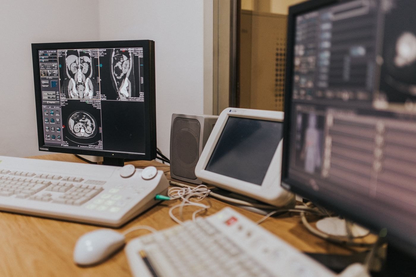
We collaborate with universities, hospitals, cardiovascular research teams, imaging laboratories and medical technology researchers. Collaboration usually means one of four things: access to the platform for a study, technical work inside a study, co-development of something new, or a feasibility exercise before a grant application.
Study design, ethics approval and data governance stay where they belong — with the institution. We contribute the technology and the people who built it.
Training is the fastest way in
Before a custom project or a platform rollout, most teams start with a workshop. It costs little, it settles quickly whether the approach helps you, and it is the same people who would do the larger work.